Persistent Müllerian Duct Syndrome in an 8-month old intact male Basset Hound
Our Case of the Month!
Pyometra with Male Genitalia 😱
An 8-month-old intact male Basset hound was scheduled for a routine neuter procedure but prior to the procedure, he presented for painfully swollen testicles.
Externally the patient had normal male genitalia with the exception of large, swollen testicles.
Internally the patient had fluid filled tubular structures resembling what appeared to be a pyometra, a surprising thing to see in male dog.
Images for this case were provided by Kelly Vazquez, CVT, SDEP® certified clinical sonographer for SonoPath Mobile Veterinary Ultrasound with interpretation by Dr. Eric Lindquist, Founder & CEO of SonoPath.com.
Excellent care and patient management by Dr. Jill Shiffman, (owner of Bergen County Veterinary Center, Waldwick, NJ) and her compassionate team.
History
The patient presented for ADR, not eating well, progressive lethargy, and swollen testicles. The owner noted that his testicles had always been large and that he does have some degree of dietary indiscretion. The patient received buprenorphine and SQ fluids pending ultrasound interpretation.
Image Interpretation
The left testicle was uniform and measured 2.64 cm. The right testicle measured 2.4 cm and was uniform. The prostate was slightly enlarged and measured 3.0 cm with minor, heterogenous parenchymal changes. The prostate revealed striating edema lines. This is consistent with prostatitis. A large amount of free fluid was noted within the testicular sac. Echogenic fluid filled tubular structures were noted in the caudal abdomen. The material in the tubular structures appeared to be echogenic and likely purulent. These encompassed the urinary bladder and were in the region of the uterus. However, no ovaries were found. Hermaphroditism should be considered. Surgical exploratory would be warranted.
DX
Outcome


Patient Information
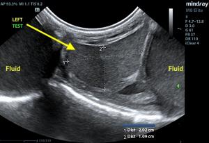
Left testicle surrounded by pockets of fluid.
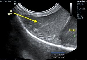
Right testicle with pocket of fluid.
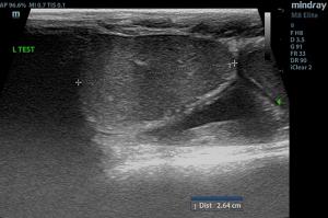
Linear image of left testicle.
.jpg)
Linear image of right testicle.
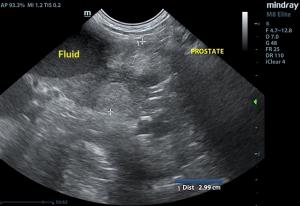
Prostate with pocket of fluid to the left,
NOT the urinary bladder.
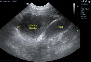
The urinary bladder was being pushed forward by
the surrounding fluid-filled structures.
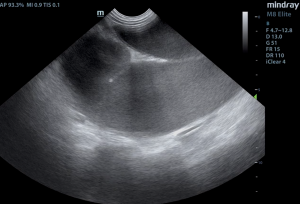
Fluid filled tubular structure.
Resources
Free Course: Reproductive Clinical Sonography
If you're struggling to achieve these views, you are not alone! The 6 lessons include Anatomy and understanding, step-by-step instructions on image acquisition, as well as hand and probe positioning. Common challenges and practical solutions are covered.
Enjoy 50% Off Any Online CE Course!
Take advantage of this HUGE discount! We have dozens of online Race approved CE courses available in a variety of topics from ultrasound, cardiology, internal medicine, dentistry, feline, and more!
Use Coupon Code: PYOMETRA
Latest Case Studies Delivered to Your Inbox - 1x Month
We highlight different cases each month that are clinically interesting, or have unusual presenting clinical signs, and/or diagnostic findings, that highlight the effectiveness of a variety of imaging modalities; ultrasound, CT, Xray.
Purchasing A New Ultrasound?
💰 Save up to $10,000 Savings with Tax code 179
🗓️ 0 Payments until 2021
🎓 Free hands-on training included
SonoPath's Mindray Elite 8 has a quick, easy-to-use workflow, and comes loaded with practical features (including cardiac software) that can grow with you, rather than limit you.
What You'll ❤️ Love about The Mindray Elite 8
✔️ Easy interface - comes with a step-by-step user guide
✔️ Efficient knobology - reduce the # of steps in your workflow
✔️ Flawless images - even on large dogs
✔️ Our support - the best in the business
Starting at $27,500, and includes a 5-year warranty.

Fostering The Art Of Veterinary Medicine®
SonoPath is a DVM owned, customer-centric company that serves a small community of veterinarians and technicians who share our passion for diagnostic efficiency, and value customer service.
We're highly focused on providing quality education, personalized support and encouragement to help our customers learn, grow, and provide the best patient care possible.
This core belief allows us to create lasting, meaningful relationships with our customers, who know they can count on us.
